CT-derived total lung volume and mortality risk in smokers with preserved ratio impaired spirometry: a lung cancer screening cohort study
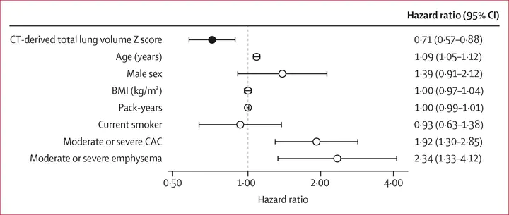 Adjusted mortality risk within PRISm according to CT-derived total lung volume (Figure 4)
Plain Language Summary
Some smokers have reduced lung function on breathing tests without the airflow blockage seen in COPD, a pattern called PRISm. People with PRISm are more likely to die early and to have heart and metabolic disease, but breathing tests are often not done in lung cancer screening. Using CT scans from over 7,500 people in the SUMMIT lung cancer screening study, we automatically measured the total size of each person’s lungs. People with PRISm tended to have smaller lungs, and among them, those with the smallest lungs were at the highest risk of dying. Because these scans are already taken for screening, lung size could be measured at no extra cost to flag people who may need breathing tests and closer follow-up.
Abstract
Background: Preserved ratio impaired spirometry (PRISm) is associated with increased mortality and cardiometabolic disease and is biologically heterogeneous. In lung cancer screening, although low-dose CT images are acquired routinely, spirometry data are often unavailable. CT-derived total lung volume could capture structural lung size directly, although its prognostic role within PRISm has not been established. We aimed to investigate whether CT-derived total lung volume identifies a high-risk PRISm phenotype and could serve as an opportunistic imaging marker. Methods: In this retrospective observational cohort study, we analysed data from SUMMIT, a prospective, UK lung cancer screening cohort. Participants aged 55–77 years were included in this study if data on baseline spirometry and automated CT-derived total lung volume measurements were available; those with missing spirometry or CT-derived total lung volume data were excluded. Demographic, smoking, spirometric, CT imaging, and comorbidity data were collected at baseline, and deaths were ascertained through linkage to national mortality records. The primary endpoint was all-cause mortality. CT-derived total lung volumes were standardised as a sex-specific Z score adjusted for body size. Multivariable logistic regression was used to evaluate associations between lung volume and PRISm and prevalent comorbidities. Cox proportional hazards models were used to assess all-cause mortality across spirometric categories (normal spirometry, PRISm, and Global Initiative for Chronic Obstructive Lung Disease grade 1, 2, and 3 or 4 airflow obstruction). We also evaluated whole-cohort discrimination of PRISm and robustness to forced vital capacity (FVC) adjustment and censoring of lung cancer-related deaths. Findings: This study included 7535 participants (mean age 65 years [SD 6]; 4503 [59·8%] males and 3032 [40·2%] females). 1355 (18·0%) participants had PRISm. Over a median of 5·0 years (IQR 4·7–5·2), mortality was higher in participants with PRISm than in those with normal spirometry (9·1% vs 4·6%; p<0·001), with the lowest CT-derived total lung volume Z score in participants with PRISm. PRISm independently predicted mortality (adjusted hazard ratio [HR] 1·91, 95% CI 1·46–2·51; p<0·001). Within the PRISm subgroup, each 1-SD increase in lung volume was associated with reduced hazard of death (adjusted HR 0·71, 95% CI 0·57–0·88; p=0·002), persisting after FVC adjustment (adjusted HR 0·76). Across the full cohort, lung volume discriminated PRISm with moderate accuracy (area under the receiver operating characteristic curve 0·73) and higher lung volume was associated with lower odds of cardiometabolic disease. Interpretation: PRISm is characterised by reduced CT-derived total lung volume and increased mortality and cardiometabolic risk. Because low-dose CT is acquired routinely in lung cancer screening, whereas spirometry frequently is not, automated total lung volume quantification could help to identify individuals for confirmatory spirometry and risk stratification from scans already obtained. External validation is required. Funding: Wellcome Trust and the National Institute for Health and Care Research.